
Ethnic-Specific Normative Data for Bony and Mucosal Nasal Dimensions Derived from CT Scans: A Cephalometric Study in a Nigerian Population 0 1
DOI:
https://doi.org/10.60787/sjhrs.vol1no1.12Keywords:
Cephalometry, Computed Tomography, Nasal Morphometry, Forensic anthropologyAbstract
Background: The morphometric dimensions of the nasal cavity are crucial for surgical planning, forensic identification, and understanding craniofacial evolution. This study aimed to establish ethnic-specific normative data for bony and mucosal nasal cavity dimensions using Computed Tomography (CT) in a Nigerian population (Hausa, Fulani, Ibra, Yoruba and Igbo ethnic groups), with a focus on anterior, middle, and posterior nasal regions.
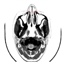
Methods: A retrospective cross-sectional study was conducted at Usmanu Danfodiyo University Teaching Hospital (UDUTH), Sokoto, Nigeria. A total of 130 head CT scans (84 males, 46 females) were analyzed. Participants (Hausa, Fulani, Ibra, Yoruba and Igbo ethnic groups) were grouped into seven 10-year intervals from 0 to 70 years. Axial CT images were used to measure: Anterior Bony Width (ABW) Right and Left Anterior Bony Widths (RABW, LABW) Middle Minimal Mucosal Width (MMW) Right and Left Middle Minimal Widths (RMMW, LMMW).
Results: The middle nasal cavity (RMMW/LMMW) reached maximum width in the 31–40 years group, with RMMW at 5.05±1.653 mm and LMMW at 4.39±1.639 mm in males. No consistent sexual dimorphism was observed across all age groups, although males generally exhibited larger dimensions. Notably, in the 31–40 years group, males had significantly greater RMMW (p = 0.007) and LMMW (p = 0.047) than females. Bilateral symmetry was preserved across all parameters (p > 0.05). The narrowest segment of the nasal cavity (middle third) was consistently < 4 mm in over 90% of subjects, indicating a high prevalence of anatomical nasal valve narrowing.
Conclusion: This study provides the first comprehensive set of ethnic-specific normative data (Hausa, Fulani, Ibra, Yoruba and Igbo ethnic groups) for nasal cavity dimensions in a Nigerian population. The findings reveal dynamic age-related changes, variable sexual dimorphism, and high bilateral symmetry. These data are essential for evidence-based surgical planning in rhinoplasty and sinus surgery, forensic anthropology, and comparative craniofacial research in African populations.
Downloads
References
1. Belden, J., Chinwuba, C., Wallman, J., & Strand, R. (1999). Nasal airway obstruction: CT assessment. Radiology, 159:503–506.
2. Berger, G., Hammel, I., Berger, R., Avraham, S., & Ophir, D. (2000). Histopathology of the Inferior Turbinate with Compensatory Hypertrophy in Patients with Deviated Nasal Septum. Laryngoscope, 110(12):2100–2105.
3. Bergstrom, L., & Owens, O. (1984). Posterior choanal atresia: a syndromal disorder. Laryngoscope, 94:1273–1276.
4. Boyan, N., Kizilkanat, E., Tekdemir, I., Soames, R., & Oguz, O. (2007). Usefulness of Nasal Morphology in Surgical Approaches for Skull Base Tumours. Neurosurg Q, 17(4):283–286.
5. Chhabra, N., & Houser, S.M. (2011). The surgical management of allergic rhinitis. Otolaryngol Clin North Am, 44(3):779–795.
6. Chinwuba, C., Wallman, J., & Strand, R. (1986). Nasal airway obstruction: CT assessment. Radiology, 159:503–506.
7. Clifford, J. B., Anthony, A. M., & Ilona, M. S. (1999). CT Features of Congenital Nasal Piriform Aperture Stenosis: Initial Experience. Radiology, 2:213.
8.Doorly, D.J., Taylor, D.J., & Schroter, R.C. (2008). Mechanics of airflow in the human nasal airways. Respir Physiol Neurobiol, 163(1-3):100–110. 9. Egeli, E., Demirci, L., Yazýcý, B., & Harputluoglu, U. (2004). Evaluation of the inferior turbinate in patients with deviated nasal septum by using computed tomography. Laryngoscope, 114(1):113– 117.
10. Ese, A.E., Okumagba, M.T., & Onodarho, E. (2013). The Facial and Nasal Height of the Ijaw Ethnic Group in Delta State of Nigeria. Advances in Applied Science Research, 4(1):1–5.
11. Evteev, A., Anikin, A., & Satanin, L. (2018). Midfacial growth patterns in males from newborn to 5 years old based on computed tomography. American Journal of Human Biology, 30(4), e23132.
12. Garandawa, H., Nwaorgu, O., & Oluwatosin, O. (2008). Morphometric Nose Parameters In Adult Nigerians. The Internet Journal of Otorhinolaryngology, 10(2):8360.
13. Ishii, L. E., Tollefson, T. T., Basura, G. J., Rosenfeld, R. M., Abramson, P. J., Chaiet, S. R., ... & Nnacheta, L. C. (2017). Clinical practice guideline: improving nasal form and function after rhinoplasty. Otolaryngology–Head and Neck Surgery, 156, S1-S30.
14. Karadag, B. et al. (2011). Racial differences in nasal bone and pyriform aperture. Aesthetic Plast Surg, 35(4):586–591.
15. Kimura, S., Chiba, R., & Capasso, R. (2013). Phase of nasal cycle during sleep tends to be associated with sleep stage. The Laryngoscope, 123(8):2050–2055.
16. Knegt-Junk, K.J., Bos, C.E., & Berkovits, R.N.P. (1988). Congenital nasal stenosis in neonates. J Laryngol Otol., 102:500–502.
17. Krishan, K. (2007). Anthropometry in Forensic Medicine and Forensic Science— 'Forensic Anthropometry'. The Internet Journal of Forensic Science, 2(1).
18. Lang, J. & Baumeister, R. (1982). Uberdas postnatale Wachstum der Nasenhöhle. Gegenbaurs Morphol Jahrb, 128:354–393.
19. Li, K.K., Powell, N.B., Riley, R.W., Troell, R.J., & Guilleminault, C. (1998). Radiofrequency volumetric tissue reduction for treatment of turbinate hypertrophy: a pilot study. Otolaryngol Head Neck Surg., 119(6):569–573. 91
20. Likus, W. et al. (2014). Nasal Region Dimensions in Children; A CT Study and Clinical Implications. BioMed Research International, 125810.
21. Marechal, L. (2023). Variation, adaptation and evolution of the nasal cavity and the nasal airway (Doctoral dissertation, Université de Bordeaux).
22. Martin, J. et al. (2008). Computer- ided Assessment of Bony Nasal Pyramid Dimensions. Arch Otolaryngol Head Neck Surg., 126(8):979–984.
23. Mohammad, W. E. et al. (2017). Computed Tomography Measurement of Inferior Turbinate in Asymptomatic Adult. Int Arch Otorhinolaryngol., 2:109–198.
24. Moore, W.J. & Lavelle, C.L.B. (1974). Growth of Facial Skeleton in Hominoidea. Academic Press.
25. Moreddu, E. et al. (2013). Morphometric Measurements and Sexual Dimorphism of the Piriform Aperture in Adults. Surg Radiol Anat., 35(10):917–924.
26. Mrig, S., Agarwal, A.K., & Passey, J.C. (2009). Preoperative computed tomographic evaluation of inferior turbinate hypertrophy and its role in deciding surgical treatment modality in patients with deviated nasal septum. Int J Morphol, 27(2):503–506.
27. Reitzen, S.D., Chung, W., & Shah, A.R. (2011). Nasal septal deviation in the pediatric and adult populations. Ear, Nose and Throat Journal, 90(3):112–115.
28. Royal, S.A., Hedlund, G.L., & Wiatrak, B.J. (1999). Single central maxillary incisor with nasal pyriform aperture stenosis—CT diagnosis prior to tooth eruption. Pediatr Radiol., 29:357–359.
29. Samadi, D.S., Shah, U.K., & Handler, S.D. (2003). Choanal atresia: a twenty-year review of medical comorbidities and surgical outcomes. Laryngoscope, 113:254– 258.
Published
Issue
Section
License

This work is licensed under a Creative Commons Attribution-NonCommercial-ShareAlike 4.0 International License.