Evaluation of some computed tomography quality control parameters in Kaduna, Nigeria 3 4
DOI:
https://doi.org/10.60787/sjhrs.vol1no1.3Keywords:
Computed tomography, Quality Control, Image NoiseAbstract
Background: Quality control (QC) tests for computed tomography (CT) are primarily concerned with the maintenance of CT scanners at the optimal operating conditions for providing the required diagnostic information at the least possible exposure to ionizing radiation. Routine QC procedures should be observed in every facility. However, an empirical review search shows no evidence of routine QC procedures being carried out in Kaduna State.
Objective: This study was aimed at evaluating some computed tomography quality control parameters in Kaduna
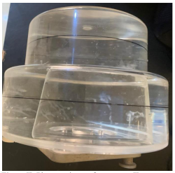
Methods: The study design was prospective and cross-sectional. Two scanners labeled Scanner I and II were included in the study. CT numbers for water, field uniformity, image noise, and high and low contrast resolution were tested using the head CT phantom
Result: CT scanners I and II passed the water and field uniformity tests (2.87 and -1.9). Both scanners failed the noise test (4.36 and 3.2). The scanner I failed the high contrast resolution test (30.55) but passed the low contrast resolution test (123.53)
Conclusion: Both scanners passed the tests for the CT number for water and field uniformity but revealed a significant failure in noise tests. The scanner passed the low contrast resolution test but failed the high contrast resolution tests. Both scanners measure tissue density reliably but excessive noise and scanner one’s poor edge definition will limit diagnostic accuracy.
Downloads
References
1. Egharevba, O. P., Nzotta, C. C., Oyekunle, E. O. & Anas, M. (2021). Assessing the status of quality control parameters of four computed tomography scanners in Ibadan. European Journal of Medical and Health Sciences, 3(4), 80–83. s
2. Jung, H. (2021). Basic Physical Principles and Clinical Applications of Computed Tomography. Progress in Medical Physics, 32(1), 1–17.
3. Seeram, E. (2016). Computed tomography: Physical principles, clinical applications, and quality control (Fourth edition). Elsevier.
4. Fred, H. L. (2004). Drawbacks and limitations of computed tomography. Texas Heart Institute Journal, 31(4), 4.
5. Ginat, D. T. & Gupta, R. (2014). Advances in computed tomography imaging technology. Annual Review of Biomedical Engineering, 16(1):431-53.
6. European commission. European Guidelines on Quality Criteria for Computed Tomography EUR 16262 EN. Luxenburg: European Commission; 1999. Available from: http//www.drs.dk/guidelines/ct/quality/page032.htm. [Last assessed on 2024 May 20].
7. Sidi, M., Hussain, U., Ya’u, A. & Garba, I. (2020). Evaluation of the computed tomography number for water, field uniformity, image noise and contrast resolutions in Kano Metropolis, Nigeria. Nigerian Journal of Basic and Clinical Sciences, 17(1), 21.
8. Dillon, C., Iii, W. B., Clements, J., Cody, D., Gress, D., Kanal, K., Kofler, J., McNitt-Gray, M. F., Norweck, J., Pfeiffer, D., Ruckdeschel, T. G., Strauss, K. J., Tomlinson, J., Davidson, C. & Hernandez, D. (2017).Computed Tomography Quality Control Manual. 86.
9. Oliveira, P. M. C., Saraiva Horta, M. A. A., Magalhaes, M. J. & Santana, P. C. (2011). Assessment of computerized tomography devices in Minas Gerais. 24-28.
10. Njiki, C. D., Ndjaka Manyol, J. E. M., Ebele Yigbedeck, Y., Abou’ou, D. W., Yimele, B. C. & Sabouang, J. F. (2018). Assessment of image quality parameters for computed tomography in the city of Yaounde.Open Journal of Radiology, 08(01), 37–44.
11. Sidi, M. (2016). Quality control tests, computed tomography scanners and Kano metropolis.Indian Journal of Applied Research, 6(3), 205-207.
12. Adejoh T, Nzotta CC, Umeh EO, Aronu ME, Dambele MY (2017). Head computed tomography protocol audit & correction in two tertiary health institutions in Anambra State of Nigeria. West African Journal of Radiology, 24(2): 157–161 DOI: 10.4103/1115-3474.206807
13. Adejoh T, Onwudiliora OE, Ukaji NF, Nzotta CC, Nwefuru OS, Aniagolu CA, Ogbonna KJ, Onuegbu CN (2015). Analysis of Artefacts From Computed Tomography of the Head at Nnamdi Azikiwe University Teaching Hospital, Nnewi. Nigerian Journal of Medical Imaging and Radiation Therapy, 4(1):6–10