Sonographic Evaluation of Renal Changes and their Correlation with CD4+ count among Human Immunodeficiency Virus Sero-positive Pregnant Women in Kano, Nigeria 1 4
DOI:
https://doi.org/10.60787/sjhrs.vol1no1.4Keywords:
HIV/AIDS, Kano, Pregnant women, UltrasonographyAbstract
Background: The prevalence of Human Immunodeficiency Virus among pregnant women in Nigeria remains a major public health challenge with serious medical, economic, and social impact.
Aim. This study was aimed at evaluating renal changes and their correlation with CD4 count among HIV sero-positive pregnant women in Kano, Nigeria.
Methods: A prospective cross-sectional work carried out from April 2021 to March 2022. A purposive sampling method was employed and included 196 patients. Mindray digital ultrasound machine with 3.5 MHz transducer was used as an instrument for data collection. The patients were scanned in supine and lateral decubitus positions. Data was analyzed using SPSS version 25.0. with a level of significance set atρ<0.05.
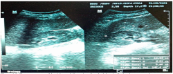
Results: The mean values of the right renal volume for the 1st, 2nd, and 3rd trimesters were 148.8 ±25.8cm3, 168.1 ±35.6cm3, and 185.2±35.8cm3 respectively whereas the left renal volumes were 163.7 ±31.1cm3, 182.4 ±40.2cm3, 199.3 ±38.4cm3. In 3rd trimester, patients had 4.55% and 1.52% increased renal parenchymal echogenicity on the right and left kidneys, respectively. A statistically significant difference in renal dimensions between trimesters was observed (ρ < 0.05). There was statistically significant weak positive correlation of right and left renal dimensions with anthropometric variables (r = 0.17- 0.25).
Conclusion: This study has established reference values for renal dimensions among HIV/AID seropositive pregnant women. A statistically significant difference in renal dimension between Trimesters was observed. The Renal dimensions had a week positive correlation with the anthropometric variables.
Downloads
References
1. Douek, D.C., Roederer, M., Koup, R.A. (2009). Emerging concepts in the immunopathogenesis of AIDS. Annu Rev Med. 60:471-84.
2. Chou, R., Cantor, A., Bougatsos, C., Zakher, B. (2012). Screening for HIV in Pregnant Women: Systematic Review to Update the U.S. Preventive Services Task Force Recommendation, Evidence Synthesis Number 96, Oregon Evidence Based practice center, Oregon, Portland, AHRQ Publication 12-05173-EF-2.
3. Szczech, L.A., Hoover, D.R., Feldman, J.G., Cohen, M.H., Gange, S.J., Gooze, L. et al. (2004). The Identification between Renal Disease and outcomes among HIV-Infected women receiving or not receiving antiretroviral therapy. Clinical Infectious Diseases. 39 (8): 1199–206.
4. Ozim, C.O., Mahendran, R., Amalan, M. Puthussery, S. (2023). Prevalence of human immunodeficiency virus (HIV) among pregnant women in Nigeria; A systematic Review and Meta Analysis. BMJ Open. 13:e050164
5. UNAIDS/WHO. Information Sheet. HIV Statistics, globally and by WHO region. 2025.
6. Adejoh, T., And Idigo, F.U. Ultrasound measurement of waist circumference & visceral adipose tissue thickness in Nigerian women. Nigerian Journal of Medical Imaging & Radiation Therapy, 2014;3(1):14-16 https://njmirt.com/issue/2014
7. Sidi, M., Ugwu, A.C., Dambatta, A.H., Saleh, M.K., Jibo, U., Jega, M.A., Aminu, A.A., Yau, A. Mansur, U. (2020). Sonographic evaluation of renal changes among drug-dependent and drug naïve adult patient with human immunodeficiency virus / acquired immunodeficiency syndrome in Kano, Nigeria, Nigerian journal of Basic & clinical sciences, 17(1):26–32.
8. Jain, R., Chopra, J., Manik, P., Sharma, P.K., Srivastava, A.K., Arora, V. (2016). Sonographic Evaluation of Renal Dimension and Their Correlation with Gender, Weight, and Height in Normal Young Adults of Uttar Pradesh Region, Indian Journal of Clinical Anatomy and Physiology, 3(2): 149–154.
9. Ugboma, E.W., Ugboma, H.A.A., Nwankwo, N.C., Okpani A.O.U. (2012). Sonographic evaluation of the renal volume in normal pregnancy at the University of Port Harcourt Teaching Hospital: A Pilot Study, Journal
of Clinical and Diagnostic Research, 6(2): 234–238.
10. Uakarn, C., Chaokromthong, K., Sintao, N. (2021). Sample Size Estimation Using Yamane, Cochran, Krejcie, Morgan, Green Formulas and Cohen Statistical Power Analysis by G-Power and Comparisons’, Apheit International Journal, 10(2): 76–88.
11. Sidi, M., Ugwu, A.C., England, A., Manafa, P.O., Dambatta A.H., Zira J.D., Saleh, M.K., Ya’u, A., and 29 The Sub-Saharan Journal of Health and Radiation Sciences | Issue 1 | Volume 1 |Jan - June 2026 Hassan et. al, Sonographic Evaluation of Renal Changes and Their Correlation with CD4+ count Among Human Immunodeficiency Virus Sero-positive Pregnant Women Mansur, U. (2021). Renal Doppler Indices and their Correlation with Laboratory Indices of Human Immunodeficiency Virus Seropositive Adult Individuals’, Radiography, Elisevier Ltd, 1–7.
12. Ugochinyere IF, Obinna AE, Chiamaka NS, Chukwunyelu AAM, Amechi AJ. (2021). Sonographic assessment of renal volume in normotensive pregnant women and women with pregnancy induced hypertension in Enugu Metropolis. Journal of Diagnostic Medical Sonography, 37(5): 442–449.
13. Eze, C.U., Adeyomoye, A. (2018). Renal echogenicity as predictor of human immunodeficiency virus associated nephropathy: a cross-sectional study at a tertiary hospital in Lagos, Southwest Nigeria. Journal of Diagnostic Medical Sonography, 34(4): 261–272.
14. Kedir, E., Berhane, M., Nigatu, T.A., Ayalew, A., Zewdu, M. (2020), Evaluation of Ultrasonic Renal Volume in Relation to Body Size in Patients with Hypertension: Comparative Cross-Sectional Study. Ethiopian Journal of Health Sciences, 34(2): 145–153.
15. Jovanovic, D., Gasic, B., Pavlovic, S. and Naumovic, R. (2013). Correlation of Kidney Size with Kidney Function and Anthropometric Parameters in Healthy Subjects and Patient with Chronic Kidney Disease. Renal Failure, 35(6): 896–900.
16. Desta, D., Zewdu, M., Ayalew, A., Nigatu, T.A. (2020). Ultrasonic Renal Size and its Correlates Among Diabetic Outpatients at Jimma University Medical Center, Southwest Ethiopia. Translational Research in Anatomy, Elsevier, 20 (100071): 1–10.
17. Zira, J.D. (2017). Sonographic Assessment of Renal Sizes, Parenchymal Thickness and Volume in Patients with Type 2 Diabetes Mellitus. Pakistan Journal of Radiology, 27(3): 213–218.
18. Akimbami, A.A., Gbadegesin, A., Ajibola, S.O., Uche, I.E., Dosunmu, A.O., Adediran, A., Sobande A. (2015). Factors influencing CD4 cell count in HIV- positive Pregnant Women in a Secondary Health Center in Lagos, Nigeria’, HIV/AIDS – Research Palliative Care, 7: 115–118.
19. Myer, L., Kamkuemah, M., Kaplancand, R. and Bekke, L.G. (2013). Low Prevalence of Renal Dysfunction in HIV-Infected Pregnant Women: Implications for Guidelines for the Prevention of Mother-To-Child Transmission of HIV. Tropical Medicine and International Health, 18(11): 1400 –1405.
20. Gwom, P.M., Atsukwei, D., Eze, E.D., Igoh, E.O., Gabkwet, E.A., Adams M.D., Rabiu, M.K., Ezekiel, I. (2021). Relationship Between Renal Ultrasonographic, CD4 Cell Count and Proteinuria Findings in HIV Infected Adult Patients in Jos, Nigeria’, African Journal of Medical and Health Sciences. 20(8): 105–118.
21. Cheung, K.L. and Lafayette, R.A. (2013). Renal Physiology of Pregnancy. Advances in Chronic Kidney Disease, 20(3): 209–214.
22. Abinaiye, P.O., Garko, S.S., Ahmed, A., Tanimu, S.S., Tahir, M.N. (2014), ‘Relationship of Ultrasound Renal Echogenicity, Serum Creatinine Level and CD4 Cell Counts in Patients with Human Immunodeficiency Virus-Associated Nephropathy’, Sub-Saharan African Journal of Medicine, 1(4): 191–197.